Histochemical Staining Methods
Organ & Disease Model-Specific Stains Available
We offer a variety of routine and special histochemicals stains that may be performed on paraffin embedded tissue sections or frozen tissue sections to visualize specific elements of interest. Our scientists are highly experienced in the optimization and application of special stains for selective staining of unique tissue elements of interest.
Below is a sampling of the types of staining methods we perform:
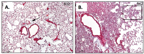
Hematoxylin and Eosin (H&E) staining
This is a classic standard tissue section staining method widely used for the inspection of tissue components for pathological analysis that’s applicable in all organs and disease models. Our scientists are well-versed in this method, based on binding of nucleic acid and other acidic components of the tissue to the basic hematoxylin stain. The acidic counter stain, eosin, binds to basic components in the tissue, such as cytoplasmic proteins.
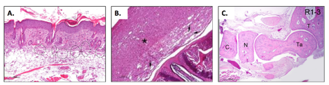
Masson Trichrome (MT) staining
This staining method is mainly used to distinguish collagen from muscle tissue, applicable and recommended in fibrosis models, wound healing studies, infarct models and others.
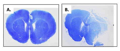
Thionine Staining
This method is used for staining of brain sections and visualizing macroscopic lesions, e.g. following MCA occlusion.
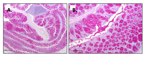
Periodic acid-Schiff (PAS) staining
This staining method is used for staining of polysaccharides such as glycogen, and mucosubstances such as glycoproteins, glycolipids and mucins in tissues e.g. colon tissues from TNBS-induced colitis model.
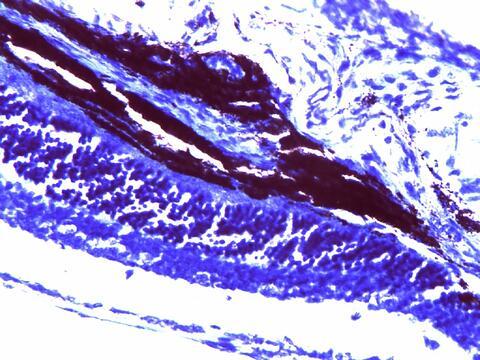
Toluidine blue staining
This method is used for staining of mast cells that are found in the connective tissue and their cytoplasm contains granules composed of heparin and histamine. Following Toluidine staining, mast cells are stained red-purple and the background is stained blue.