Digital Pathology
Precision Imaging, Pathologist Review and Quantitative Image Analysis
MLM offers cutting-edge digital pathology imaging tools to provide rapid, high-resolution digitization of whole slides for review and assessment through our secure, cloud-based image management systems (IMS).
Our experienced teams of histologists, scientists, and imaging technicians ensure sponsor samples are optimally prepared for downstream assessments by automated image analysis and/or our network of consulting pathologists.
Whole Slide Imaging (WSI)
Our systems capture high-resolution whole slides images via tiled acquisition and image stitching.
We offer:
- Scanning with 1.25x, 5x, 10x, 20x, and 40x objectives
- Brightfield imaging
- Fluorescent whole slide imaging with up to 5 fluorescent channels + DAPI counterstain
We have methods in place to standardize imaging across samples and minimize autofluorescence, channel bleed-through, and fluorescent artifacts.
Cloud-based image management systems (IMS)
Our cloud-based image management system is accessible from any internet browser without specialized software. Here, we host client images allowing easy viewing of images with large file sizes (up to 6GB+) and navigation at multiple magnifications. The system allows MLM and client teams to create and share image annotations, snapshots, and regions of interest. It also has a highly flexible database structure for field creation, data input, and reporting. Images can be shared via share links (which can be password protected if desired) and whole slide images or snapshots can be downloaded from this interface. Or, images can be archived there. For sponsors that have in-house whole slide imaging capabilities, we also provide remote uploading of most major whole slide image formats for remote review and analysis by our teams.
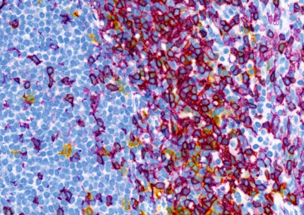
AI-powered quantitative image analysis
We provide extensive experience in quantitative image analysis to address challenging histopathology questions in an unbiased fashion with standard and customizable readouts. Analyses can be used to quantify many things in an area-based, object-based of cell-based fashion. This includes the area and intensity of staining or the percent of cells positive. Digital H-scoring can be performed to reflecting the number and intensity cells positive.
Additionally, morphometric, co-localization, and staining localization (nuclear/membranous/cytoplasmic) analyses can be applied to characterize staining. We frequently design custom read outs for specific objectives, different markers, staining localizations, and tissue types. Our image analysis platform is compatible with our cloud-based image management system such that layer data and results generated from image analysis can be uploaded and viewed online. Digital image analysis allows assessment of parameters that would otherwise be impossible to assess in glass slides and can be very complementary to manual review of physical slides by a qualified pathologist done in parallel.
Pathologist assessment
Our highly experienced board-certified pathologists partner with you to offer:
- Histopathology slide review and disease scoring
- Telepathology consultations with remote reviewing
- Assay development and consultation
- Standard and custom qualitative, semi-quantitative, and quantitative assessments
Together, our digital pathology systems can provide a complete solution to achieve your project goals.